

 With small incision sites and direct access to most areas of the joint, an arthroscopic surgeon can diagnose and surgically correct a vast array of joint problems such as arthritis and ligament tears.
In the past, treatment of orthopedic injuries involved extensive surgery, including large incisions, a hospital stay, and a prolonged recovery period. But today, with the help of an arthroscope, today’s orthopedic surgeon can easily examine, diagnose, and treat problems in the joint that previously may have been difficult to identify.
The arthroscope is a small fiber-optic viewing instrument made up of a tiny lens, light source and video camera. The surgical instruments used in arthroscopic surgery are very small (only 3 or 4 mm in diameter), but appear much larger when viewed through an arthroscope.
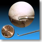
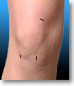 Shown at right — both as it appears on the operating table and when viewed arthroscopically — is a probe, used for examination of internal structures (in this case the underside of a patella, or kneecap). The surgeon inserts the arthroscope into the joint through a tiny incision (about 1/4 of an inch) called a portal. Two or three incisions may be made for portals. Other portals are used for the insertion of surgical instruments, such as the probe shown above. Typical incision sites and sizes for knee arthroscopy are shown at left. These incisions result in very small scars which in many cases are unnoticeable.
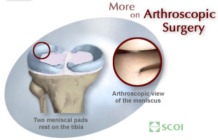
For example, almost any region of the knee may be treated arthroscopically. A normal meniscus — shown above — appears through the arthroscope appears as a smooth, white wedge-shaped structure. This QuickTime movie shows a brief portion of an arthroscopic meniscal repair.
Arthroscopic surgery is not limited to the knee: also common is arthroscopy of the shoulder, ankle, wrist, elbow, and hip. There’s even arthroscopy of the Great Toe!
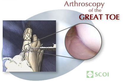 Believe it or not, the arthroscope may be used to repair damaged articular cartilage on the surface of the great toe. To the left is what the setup looks like in the operating room. The toe is held in place, and the arthroscope is inserted into the great toe joint (also inserted is a shaving instrument). To the right is the view from the arthroscope. The toe bone actually looks like it is in pretty good shape – it’s nice and smooth and shiny with no pock marks or loose bodies in there.
— Andrew M. Blecher, MD